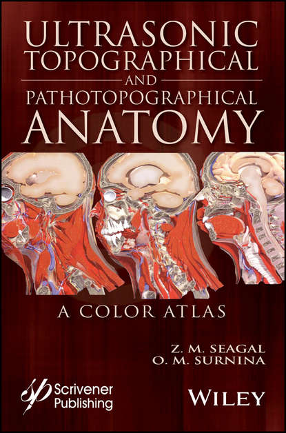
Новая книга полностью в цвете и написана опытными и уважаемыми врачами и профессорами. Она представляет собой атлас ультразвуковой топографической и патотопографической анатомии тела, включая голову, шею, грудную клетку, переднебоковую брюшную стенку, органы брюшной полости, заднебрюшное пространство, мужской и женский тазы и нижние конечности. В книге представлены конкретные и неконкретные ультразвуковые симптомы для нормальной и аномальной развивающейся анатомии, а также для диффузной и локальной патотопографической анатомии. Атлас содержит сравнительные топографические и патотопографические данные и является первым пособием такого рода для студентов и медицинских специалистов различных областей, включая специалистов медицинской сонографии. Оригинальная технология была протестирована в клиниках на пациентах, проходивших ультразвуковое наблюдение. Благодаря раннему обнаружению не было ложно-положительных или ложно-отрицательных результатов. Терапия была эффективной, а в некоторых случаях использование оригинального метода "сигалографии" (оптометрии и пульсометрии) позволило разработать новые методы лечения и/или определить оптимальные дозы лекарств, а также разработать эффективные лекарственные комплексы для лечения данной патологии. Эта важная новая книга будет полезна врачам, молодым врачам, резидентам, преподавателям медицины и студентам медицинских вузов, как учебник или справочное пособие. Она является необходимым элементом библиотеки любого врача.
Written by experienced and respected physicians and professors and profusely illustrated with the newest, cutting edge color photographs, this book offers the new clinically validated subjects of ultrasonic topography and pathopatology of the human body-head, neck, thorax, antero-lateral abdominal wall and abdomen, retro-parietal space, pelvic girdles and both male and female lower limbs in distinct separate chapters. Encyclopedic descriptions of each region are inter-spersed with illustrated black and white anatomical diagrams. Major symbols indicate relevant consulted anatomy books (e.g. Grossman 1992; Gray's 38th ed.; Netter 2008). Accurate details highlight key features which differentiate normal anatomy from abnormal variants of both development and disease along with descriptive tabular summaries of normal physique versus pertinent relevant disease states. There are specific suggestions offered for both specific and nonspecific ultrasonic symptoms associated with either normal or abnormal developing variants, involving either diffuse or localized pathophatological conditions. This comprehensive volume not only proves to be an informative orthopedic guide but an invaluable resource in topography, anatomy, medicine both for graduate students, physicians of many different specialty fields both in practice and academia, demonstrating features contrasting one topography with another and how these relate to various pathological entities.
Электронная Книга «Ultrasonic Topographical and Pathotopographical Anatomy» написана автором Z. M. Seagal в году.
Минимальный возраст читателя: 0
Язык: Английский
ISBN: 9781119224044
Описание книги от Z. M. Seagal
Written by experienced and well-respected physicians and professors, this new all-color volume presents the ultrasonic topographical and pathotopographical anatomy of the body, including the head, neck, chest, anterolateral abdominal wall, abdominal organs, retroperitoneal space, male and female pelvises, and lower extremities. Specific and non-specific ultrasonic symptoms are suggested for normal and abnormal developmental variants, diffuse and local pathotopographical anatomy. This color atlas contains comparative topographical and pathotopographical data and is the first manual of its kind for students and medical specialists in different areas, including those specializing in medical sonography. The original technology was tested at clinics in patients subjected to ultrasonic monitoring. Because of early detection there were no false-positive or false-negative results. The therapy was effective, and, in some cases, the use of the original method of “seagalography” (optometry and pulsemotorgraphy) has made it possible to develop new methods of treatment and/or to determine the optimal doses of drugs, as well as to develop effective drug complexes for treatment of a given pathology. This important new volume will be valuable to physicians, junior physicians, medical residents, lecturers in medicine, and medical students alike, either as a textbook or as a reference. It is a must-have for any physician’s library.
